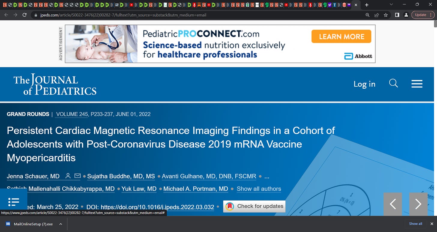
DEVASTATING!: "Persistent Cardiac Magnetic Resonance Imaging Findings in a Cohort of Adolescents with Post-Coronavirus Disease 2019 mRNA Vaccine Myopericarditis" (Schauer et al.); 'cardiac magnetic
by Paul Alexander
resonance imaging findings 16 patients, aged 12-17 yrs, with myopericarditis after 2nd dose of Pfizer mRNA vaccine. many had persistent cardiac magnetic resonance imaging findings 3-8 months post shot
This means that the effect from spike on heart myocardia is long-term, longer-term, and not transient, myocarditis is not ‘mild’. It can be catastrophic.
‘We describe the evolution of cardiac magnetic resonance imaging findings in 16 patients, aged 12-17 years, with myopericarditis after the second dose of the Pfizer mRNA coronavirus disease 2019 vaccine. Although all patients showed rapid clinical improvement, many had persistent cardiac magnetic resonance imaging findings at 3- to 8-month follow-up.
This group had a median age of 15 years (range, 12-17 years), were mostly male (n = 15, 94%), White, and non-Hispanic (n = 14, 88%). One patient was Asian, and 1 patient was American Indian. Median time to presentation from the second dose of the Pfizer COVID-19 mRNA vaccine was 3 days (range 2-4 days). All patients had chest pain. The most common other presenting symptoms were fever (n = 6, 37.5%) and shortness of breath (n = 6, 37.5%). All patients had elevated serum troponin levels (median 9.15 ng/mL, range 0.65-18.5, normal <0.05 ng/mL). Twelve patients had C-reactive protein measured with median value 3.45 mg/dL, range 0-6.5 mg/dL, normal <0.08 mg/dL.
Ten (62.5%) patients had an abnormal ECG, with the most common finding being diffuse ST-segment elevation.
SOURCE:
https://www.jpeds.com/article/S0022-3476(22)00282-7/fulltext?utm_source=substack&utm_medium=email
A total of 35 patients with the diagnosis of myopericarditis associated with Pfizer COVID-19 mRNA vaccine were followed at our institution. Twelve patients were excluded, as they never had cardiac MRI scans due to delayed presentation after initial symptoms resolved or admission to other centers. Six patients were excluded, as they did not have a follow up cardiac MRI, either because they followed up out of state or a study is still pending. One patient was excluded, as initial cardiac MRI was performed 3 weeks after presentation. Sixteen patients who had both acute-phase and follow-up cardiac MRIs available for review comprised the final cohort. This group had a median age of 15 years (range, 12-17 years), were mostly male (n = 15, 94%), White, and non-Hispanic (n = 14, 88%).’

