Polykretis: "Role of the antigen presentation process in the immunization mechanism of the genetic vaccines against COVID-19 and the need for biodistribution evaluations"
by Paul Alexander
This paper gives a very decent background primer into the process via which initial immune response happens at the cellular level after the mRNA is translated by the ribosomes and RNA polymerases etc.
The mechanism of ‘traditional’ vaccines consists in inoculating viruses, which have been previously inactivated (e.g. by thermal treatments), or attenuated (e.g. by multiple passages in suboptimal growth conditions).1 Such viruses, which lost the ability to cause acute infection, allow the immune system to recognize them as exogenous pathogens, promoting the production of specific antibodies and memory-T lymphocytes.
SOURCE:
https://onlinelibrary.wiley.com/doi/10.1111/sji.13160
‘The mechanism of ‘traditional’ vaccines consists in inoculating viruses, which have been previously inactivated (e.g. by thermal treatments), or attenuated (e.g. by multiple passages in suboptimal growth conditions).1 Such viruses, which lost the ability to cause acute infection, allow the immune system to recognize them as exogenous pathogens, promoting the production of specific antibodies and memory-T lymphocytes.1 The genetic vaccines against COVID-19 which obtained the authorization for use in the European Union, namely the adenoviral-based vaccines (produced by AstraZeneca and Janssen) and the mRNA vaccines (produced by Pfizer/BioNTech and Moderna), encode genetic information, which enables human cells to produce a viral antigen. More precisely, the aforementioned vaccines induce the protein synthesis machinery of human cells to translate the spike protein of the viral capsid of SARS-CoV-2.2
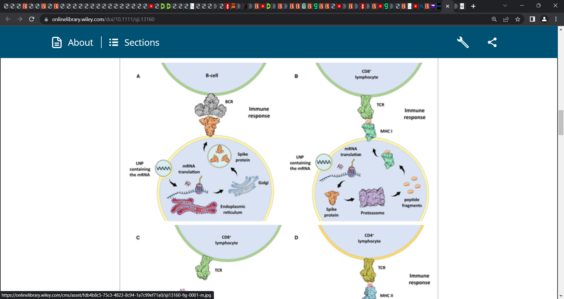
Upon its translation by the ribosomes, the spike protein gets processed by the Golgi apparatus and presented to the immune system in two forms: i) as an entire protein, displayed on the cellular membrane, which can be recognized by B cells and T-helper cells (Figure 1A); or ii) in the form of fragments loaded on the major histocompatibility complex I (MHC I), which presents the endogenous antigens to CD8+ T lymphocytes (Figure 1B).
The immune system recognizes the exogenous antigen, initiates the inflammatory response and the subsequent steps leading to the production of specific antibodies by the B cells.2 In human cells, the antigen presentation process is performed by the MHC I and II, and this mechanism is essential for the cell-mediated immunity.3 The MHC I is a protein complex, located on the membrane of all nucleated cells, which presents to CD8+ lymphocytes fragments of endogenous antigens, generated upon the proteasomal degradation of intracellular proteins (Figure 1C).3 This mechanism allows the immune system to constantly screen the proteosynthetic activity of all nucleated cells of the body, in order to detect when a cell is synthesizing viral or mutant proteins.
The MHC II is located on the membranes of professional antigen-presenting cells (APCs), such as macrophages, monocytes, B cells and dendritic cells, and it displays fragments of exogenous antigens ingested around the body to CD4+ lymphocytes (Figure 1D).3 In some cases, MHC II molecules can be found even on endothelial cells, as a consequence of inflammatory signals.3 When a CD8+ or CD4+ lymphocyte detects a cell expressing a viral gene (e.g. due to an infection), a mutant gene (e.g. due to cancer) or a foreign gene (e.g. due to a transplant), it binds the MHC activating the immune response that leads to the destruction of the abnormal cell.3
The aforementioned processes are essential for understanding the differences between the ‘traditional’ and the genetic vaccines, in terms of antigen presentation. The ‘traditional’ vaccines generally do not induce human cells to produce viral proteins, and thus, human cells do not expose viral antigens deriving from their proteosynthetic activity. On the contrary, the genetic vaccines against COVID-19 induce human cells to produce the spike protein, relying intrinsically to an autoimmune reaction, extended to all the cells that intake the genetic material and start the protein synthesis.’